|
e-mail:
jgrossh@gwdg.de |
|
Nuclear morphology - stem cells - ageing
|
|
In many differentiation processes the nuclear shape changes dramatically
(e. g. neutrophil granulocytes). During cellularisation in Drosophila the
shape of the nuclei changes from spherical to ellipsoid and the length
increases by a factor of 2.5. By mutant analysis, we identified the novel
farnesylated lamina protein Kugelkern (kuk).Kuk
is required for the nuclear shape change and encodes a novel
farnesylated protein that
localises to the inner nuclear membrane. Similar as lamin B,
Kugelkern is farnesylated and induces overproliferation of nuclear
surface area and ruffled and lobulated nuclei when overexpressed.
It is unclear how Kuk and LamB affect the morphology of the nuclear envelope and
how it mediates the interaction with chromatin.
The resulting
nuclear morphology is reminiscent to nuclear morphology in cells from patients
suffering from the Hutchinson-Gilford progeria syndrome which is caused by a
dominant mutation in the laminA gene.
To reveal the molecular mechanism of accelerated ageing in HGPS, we established a Drosophila
model for HGPS by tissue specific expression of farnesylated Lamin or Kugelkern (Brandt 2008).
Currently, we test the hypothesis that the LINC complex mediates the ageing inducing effect of
farnesylated LaminA (Chen, Cell 149 (2012)). In comparison to vertebrates, the genetic
complexity of the linc complex is much lower, with only one SUN protein (klaroid) and two
KASH protein (Klarsicht, MSP300).This simple gene structure avoids problems of genetic
redundancy.
One of the current models for the mechanism of HGPS is the stem cell depletion model
(e. g. Espada J Cell Biol 2008). It was
proposed that the altered structure of the nuclear lamina changes the proliferation and
differentiation behaviour of stem cells and that this ultimately would result in either loss
of stem cells or mis-differentiation. To test this model we apply our HGPS model onto
the intestinal stem cells of adult flies.
(see Brandt 2006, Brandt 2008, Polychronidou 2010, funding:
Niedersächsisch-Israelische Forschungsprojekte (VW Stiftung), Dorothea-Schlözer programme)
|
|
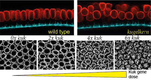
Nuclei elongate from a sperical shape with 4 µm diameter to an ellipsoid with a
length of 10 µm during cellularisation. In kugelkern mutants the nuclei remain sperical (red: nuclear
envelope stained for Lamin, blue: staging by staining of the furrow canal by Slam). The gene dose of kugelkern
determines the nuclear surface area. Embryos with extra gene copies of kuk (4x, 6x) show a ruffled shape in
cross-sections.
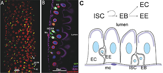
Cell types in mid-gut of adult Drosophila. (A) surface view of midgut epithelium stained for
nuclear envelope/LaminDmO (red), GFP reporter for stem cells+enterblasts (green). (B) Confocal section of
gut epithelium stained for nuclear envelope/LaminDmO (red), DNA (blue),GFP reporter for stem
cells+enterblasts (green), F-actin (purple). (C) Scheme of differentiation pathways. Scheme of gut
epithelium. Cell types: intestinal stem cells (ISC, enteroblasts (EB), enterocytes (EC), endocrinic cells
(EE), visceral muscle (mc).
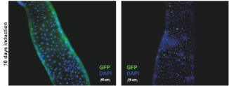
Flip-out clones induced in stem cells after 10 days expressing GFP (left panel) and GFP+LaminDmO
(right panel). Note that expression of LaminDmO suppresses proliferation of clonal cells.
|
|