Mouse hippocampal glial cell (organotypic slice) expressing the optical redox-sensor roGFP1
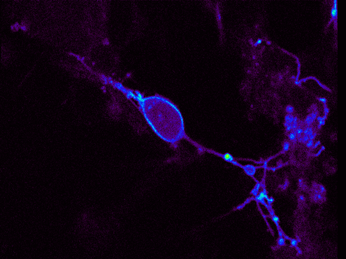
Cultured rat hippocampal neuron expressing the voltage-sensitive optical probe hVOS. Fluorescence donor of this hybrid FRET construct is a membrane-anchored eGFP
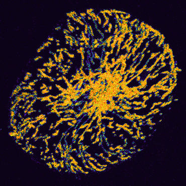
Cultured rat hippocampal astrocyte labeled with JC-1. The ratio of red and green JC-1 fluorescence, reports the mitochondrial polarization. Red-yellow colors indicate a high degree of polarization, blue/green colors represent weak polarization
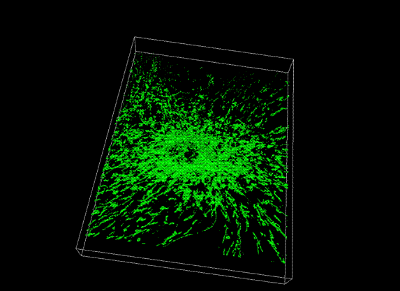
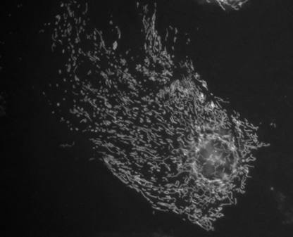
3-dimensional reconstruction of Rh123-labeled mitochondria in a cultured hippocampal astrocyte
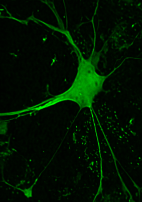
Mouse hippocampal astrocyte (cultured) transfected with the mitochondrially-targeted optical H2O2 sensor HyPer
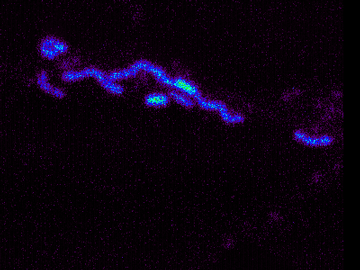
Individual Rh123-labeled mitochondria within the dendrite of a cultured brainstem neuron
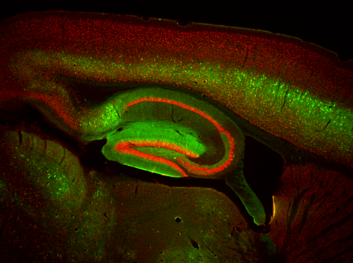
Sagittal slice of a mitochondrial roGFP1 (green) expressing mouse counterlabeled with the neuronal marker NeuN (red)
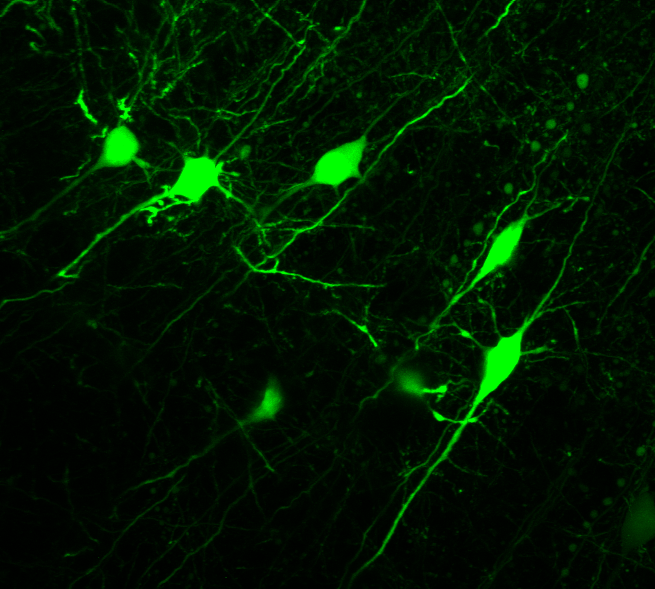
Cortical neurons expressing cytosolic roGFP1 (acute brain slice, p10)
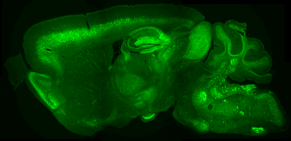
Sagittal brain slice of a transgenic mouse expressing the mitochondria-targeted redox-sensor roGFP1. Image is composed of several individual CCD camera images