X-ray Computertomography Investigation of Head and Thorax of Priacma serrata (Coleoptera: Archostemata) |
Method |
|
The specimens for the X-ray tomography were transferred to 100% ethanol and dried at the critical point. The dry specimens were mounted with soft wax to the sample stage of the x-ray machine. |
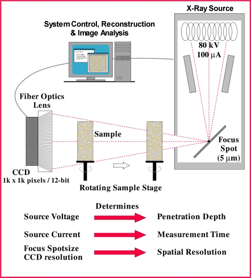
| All measurements and reconstructions were made with a Skyscan 1072 high-resolution Micro-CT system. MicroCT (Computerised Tomography) is a method for non-destructive 3D X-ray microscopy. The basic principles of the technique are similar to those used in medical CT-scanners. Recent improvements in the development of X-ray sources and CCD-detectors now make it possible to achieve a resolution down to a few microns. | |
| The Skyscan Micro-CT system consists of a sealed microfocus X-ray tube (80 kV, 100mA, 5mm spotsize), a fiber-optics coupled, cooled CCD-detector (field of view 22x22 mm, 1024x1024 pixels, 12-bit A/D conversion), a precision object manipulator and a dual-processor computer for system control and reconstruction of cross-sections. |
|
The measurement is performed by placing the object on the specimen stage, in between the X-ray source and the CCD detector. As the source produces a cone beam of X-rays, magnification is achieved by moving the object towards the X-ray source. After positioning, 2D X-ray images (radiography) are acquired from 400 angles over 180 degrees (total acquisition time about 30 minutes), and saved in the computer memory. Now cross-sections of the object are reconstructed by applying the “filtered back-projection algorithm for fan-beam geometries”. This algorithm combines all angular information for every line in the camera to generate a cross-section of the object (reconstruction time about 20 seconds per cross-section). With this method one can reconstruct up to 1024 (size of the CCD detector) cross-sections of the object, with a lateral resolution, and a slice-to-slice distance, down to a few microns. For the dataset used for this study the pixel size is 5.4µm and the cross-section to cross-section distance is 10.8µm. The magnification is 44 times. |
X-ray projections: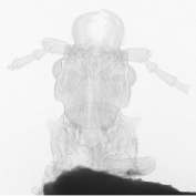 Head and prothorax. |
 Meso- and metathorax |
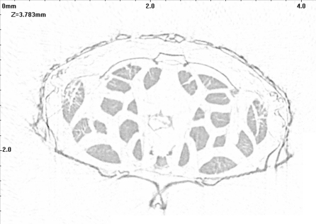
| Reconstructed crossection of thorax: | |
|
|
|
|
Greyscale represents X-ray absorption |
Flase color representation |
| The 3D
reconstruction and analysis of the X-ray data was done with the Research
Systems NOESYS software package on a dual Pentium III 550 MHz computer. The
NOESYS software allows the recombination of the X-ray sections into a
three-dimensional model of the object.
This model can be manipulated in different ways. It is possible to rotate the model around all three axes and to change the magnification. One can also remove parts of the dataset to look into the beetle. Since the X-ray data only represent absorption values it is necessary to apply “false” colors to the dataset to produce greyscale or color images. Via the color-tables it is also possible to change the opacity of selected colors or absorption values. |
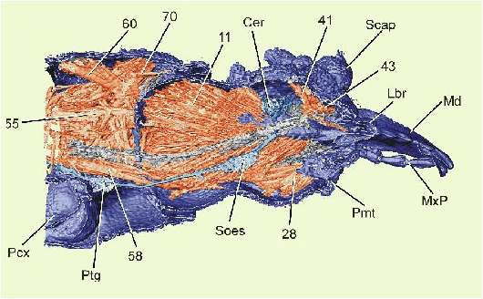
Sagittal section of meso- and metathorax: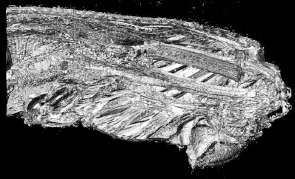 in greyscale and |
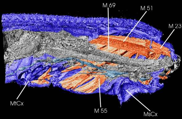 in false color representation (M51, 55, 69 = muscles of metathorax; M23 = dorsal longitudinal muscle of mesothorax; MtCx, MsCx = meta- and mesocoxa; in grey = intestine; in light blue = ventral nerve cord with ganglia. Nomenclature of muscles from Baehr (1975)) (click image to see bigger version) |
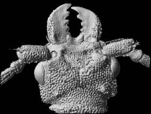
| Ventral view of head | |
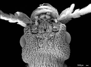 SEM image |
 reconstructed from X-ray data |
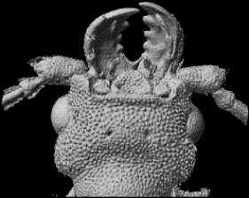
| Dorsal view of head: | |
 reconstructed from X-ray data |
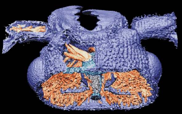 Head seen from behind and above, with dorsal cut-out to show antennal muscles. (click image to see bigger version) |
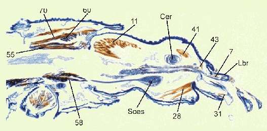
| Sagittal section of head and prothorax: | |

Numbers indicate muscles (nomenclature after Kéler
(1963)); Cer = Cerebrum; Lbr = Labrum; Md = Mandibel; MxP = Palp of
maxilla; Pcx = Procoxa; Pmt = Prementum; Ptg = Ganglion of prothorax; Soes
= Suboesophageal ganglion. |
 Histological section
|
ReferencesBaehr M. 1975. Skelett und Muskulatur des Thorax von Priacma serrata Leconte (Col.: Cupedidae). Z Morph Tiere 81:55-101. Hörnschemeyer T, Beutel RG, Pasop F. 2002. Head Structures of Priacma serrata Leconte (Coleoptera, Archostemata) Inferred from X-ray Tomography. J. Morphol. 252:298-314. Kéler S v. 1963. Entomologisches Wörterbuch. Berlin: Akademie Verlag. 774 p. |