|
|
|
Mitochondrial organization and motility |
|
|
|
 |
|
|
|
This projects aims to unveal the mechanisms controlling mitochondrial organization and motility as well as the functional implications of mitochondrial (re-)arrangement in view of cellular physiology and neuronal responsiveness
|
|
|
|
|
|
|
 |
|
|
 |
|
|
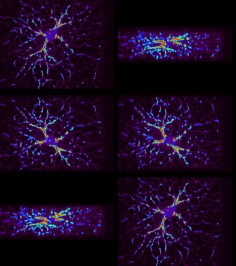
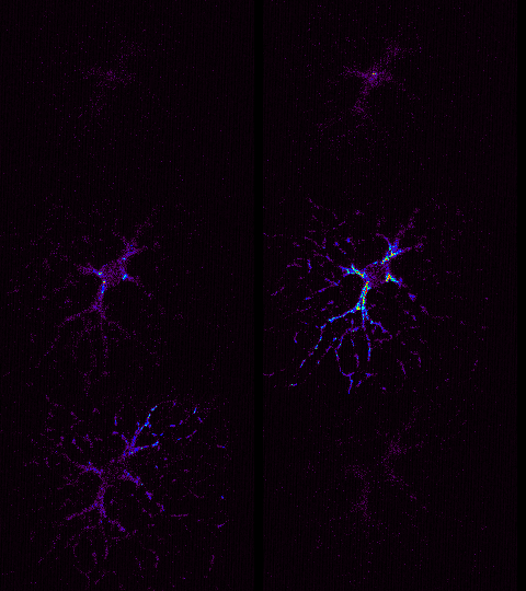
| Mitochondria form a well organized network in cultured respiratory neurons, which consists of individual mitochondria, filamentous structures and irregular-shaped clusters. This network is analyzed using the unique advantages of 2-photon laser scanning microscopy. For visualization mitochondria are fluorescently labeled, either by the mitochondria-specific marker rhodamine123 or by transfection with cyan-fluorescent proteins coding cytochrome oxidase. |
|
|
|
3-D reconstruction of the Rh123-labeled mitochondrial structures
|
|
|
|
Optical sections (XY-planes) of the mitochondrial network
|
|
|
|
 |
|
|
|
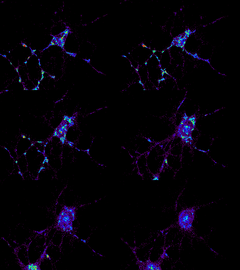
The organization of the mitochondrial network is highly dynamic. Single mitochondrial structures undergo both, Brownian motion as well as directed movements. The latter type of movement apparently occurs along microtubules, possibly by involvement of molecular motors. Disruption of microtubules by colchicin results within minutes in the breakdown of the mitochondrial network and the subsequent accumulation of mitochondria in the swelling soma. |
|
|
|
Breakdown of the mitochondrial network upon disruption of microtubules
|
|
|
 |
|
|
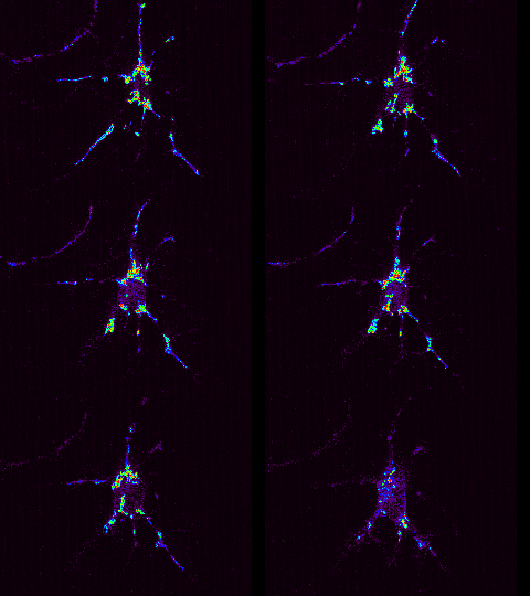
| Forskolin and IBMX reversibly arrest mitochondrial motility, suggesting that protein phosporylation downstream of cAMP may be involved in the transition from motile to resting mitochondria. |
|
|
|
Forskolin-induced arrest of mitochondrial motility
|
|
|
|
|
|
|
|
|
|
|
|
|
|
|
|
|
|
|
|
|
|
|
|
|


