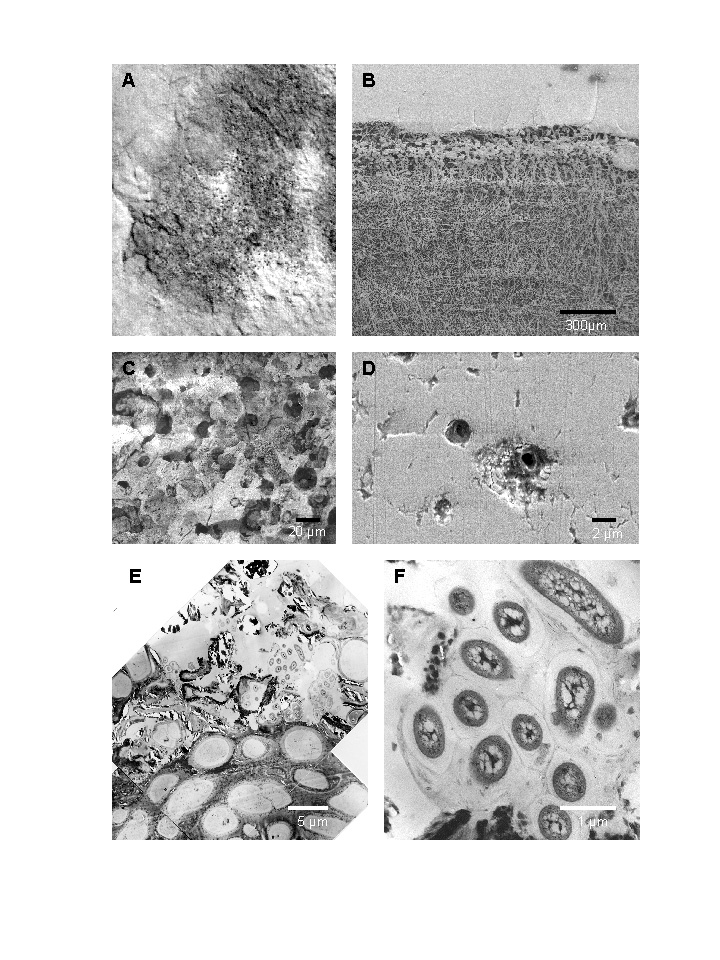
A) Small scale mosaic of endolithic lichens on carbonate rocks (the
figure shows a sector of apprx. 5 x 4 cm original size).
(Dachstein, Austria)
B) Typical structure of a sample taken from a respective site visualized
by scanning electron microscopy (SEM). The section was decalcified by incubation
in organic acids before preparation for SEM. The carbonate substratum is
completely removed. The upper layer is dominated by algal cells (photobiont),
the large lower layer consists of with fungal hyphae.
C) Photobiont layer in an untreated sample. The small cavities are
created by active substrate dissolution of the algae.
D) Detail of hyphae dissolving the limestone. Scanning electron micrographs
of sections perpendicular to the rock surface.
E) Transmission electron micrographs of the upper layer in an endolithic
biofilm, consisting of the fungal mycelium, an upper layer of photobionts
and microcolonies of heterotrophic bacteria (F depicts an enlarged view).