Research group of Jörg Großhans

Institute of Developmental Biochemistry
Universitätsmedizin
Georg-August-Universität Göttingen


home

research

people

publications

jobs

links

gallery

contact

e-mail:

jgrossh@gwdg.de


NEWS:

2.10.19 Anja and Zhiyi are moving to Chapel Hill and Qingdao. We wish them all the best.


2.10.19 Deqing's CaLM paper has been published in EMBO reports


30.9.19 Boyang's cell cycle papers are out in CELL CYCLE and G3


6.2.19 The SHMT/dNTP paper is in press with Current Biology


Structure formation in early Drosophila embryos


Research topics


Epithelial morphogenesis and cell cycle switch

- Membrane and epithelial compartment formation
- Actin dynamics and Rho signalling
- Role of the slam RNA-protein complex
- Cell cycle control


Quantitative morphogenesis (Biomechanics)

- Nuclear ordering and cytoskeletal network dynamics
- Cell intercalation and junction dynamics


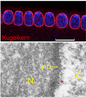
Size and form of the nucleus - Ageing

- Farnesylated nuclear lamina proteins
- Drosophila model for the Hutchinson-Gilford progeria (HGPS) and other laminopathies
- Lamina proteins in regeneration and proliferation of intestinal stem cells


Methods


- Drosophila genetics, embryology, adult ageing
- Molecular genetics
- Biochemistry
- Classical and quantitative microscopy and live imaging
- Micro-surgery with UV laser
- Computational image analysis

Cross-section of an embryo during early gastrulation. Adherens junctions (red), RhoGEF2 (green), DNA (blue). Ventral cells show specific cell behaviour, in that they change their shape initiated by apical constriction triggered by relocalisation of RhoGEF2 from basal to apical.

kugelkern mutant embryos do not elongate their nuclei during cellularisation, leaving them sperical. Consitent with its function the Kugelkern protein is associated with the inner nuclear membrane similar to lamina proteins. Similar to LaminDmO Kugelkern is farnesylated at its C-terminus what promotes membrane association. Immunofluorescence and immuno-electron microscopy in which Kugelkern is marked by 10 nm Gold particles.